
‹
›
‹
›
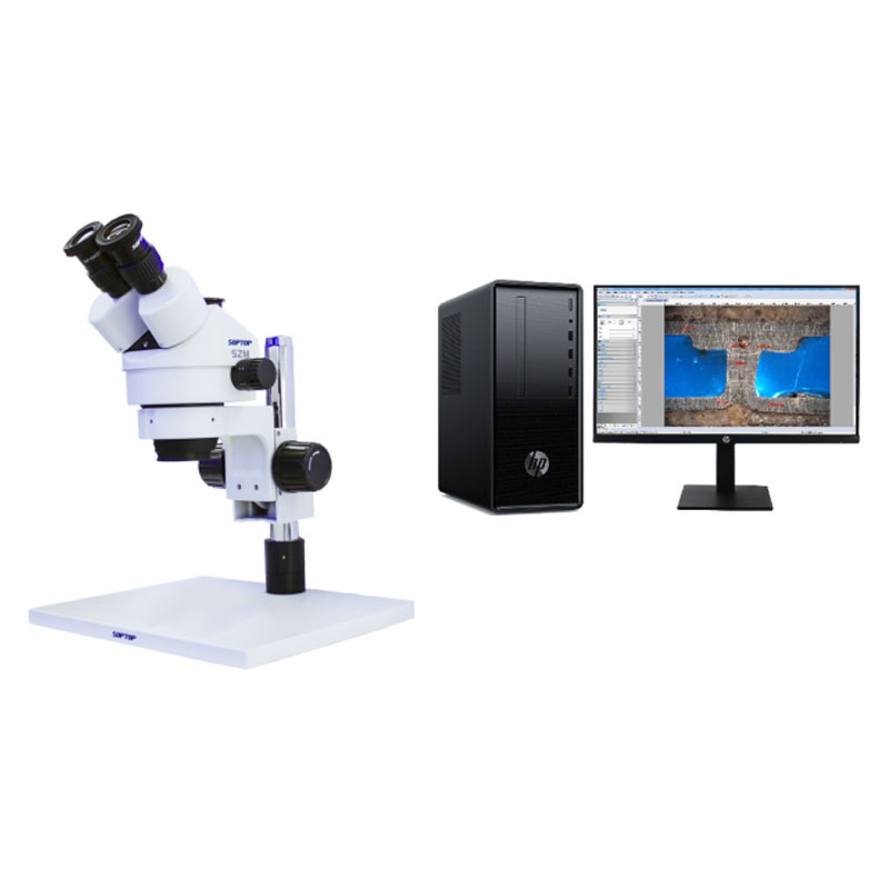
SZM-W Computerized Continuous Zoom Stereo Microscope
Overview of the SZM-W Computerized Stereo Microscope
The SZM-W Computerized Stereo Microscope is a trinocular continuous zoom stereo microscope. Its classic design ensures superior body stability, while its excellent optical performance, complete accessories, and diverse combination configurations meet the online inspection needs of modern biology, medicine, scientific research, and the modern electronic industry.
High-Quality Optical System
Optical components coated with special films create a high-performance optical system.
Wider Magnification Range
Equipped with a continuous zoom objective lens of 0.7X~4.5X (6.4:1), the standard magnification ranges from 7X to 45X. With optional eyepieces and auxiliary objective lenses, a magnification of 3.5X-90X can be achieved.
Extra-Long Effective Working Distance
The effective standard working distance reaches 100mm. With optional auxiliary objective lenses, the working distance can be extended to 26mm~287mm, providing sufficient space for operation.
Configuration Table of the SZM-W Computerized Stereo Microscope
Note: "" is standard; "O" is selected
FEG Series Digital Microscope Camera
Innovative technology significantly boosts the USB 2.0 frame rate while ensuring raw image output, making it the preferred choice for simple and cost-effective microscopic imaging.
FCL-RS Metallographic Image Measurement Software

Overview
FCL-RS Metallographic Image Measurement Software is the basic version of image measurement software launched by our company, supporting multiple developable functions. In addition to basic capture and measurement capabilities, it includes automatic panoramic image stitching, video EDF (Extended Depth of Field) extension, video overlay of time and scale bars, accurate layer fusion of measurement data and images, and support for saving in various image file formats.
Features
l Real-time static image capture: Capture static images as per user requirements.
l Multi-format image saving: Save images in JPG, BMP, PNG, and TIF formats.
l Fast and intuitive calibration: Calibrate using scale lines for precise measurement foundation.
l Diversified image processing: Support processing for the entire image or specific target regions.
l Advanced measurement functions: Calculate key geometric parameters including perimeter, width, radius, circumference, and angle.
l Real-time dynamic stitching: Achieve panoramic sample shooting and expand the visible field of view effectively.
l Multi-depth image fusion: Merge images captured at different depths of field into a single high-definition clear image.
Basic Functions
1. Image Shooting
Support real-time static image capture, batch image collection and saving, as well as batch addition of scale bars and other annotation texts.
2. Geometric Measurement
Provide a variety of measurement tools including distance, rectangle, circle, polygon, polyline length, angle, straight line included angle, radian, and point-to-center distance to meet users' basic geometric measurement needs and obtain relevant measurement data.
Measured data can be automatically fused with images, intuitively displaying the accuracy of the measured positions.
3. Dynamic Stitching
For customers requiring inspection of a larger field of view, the software is equipped with an image stitching function. Users can move the microscope's X-Y platform to meet the demand for capturing images with an expanded field of view, resolving the inconvenience of being unable to take complete shots due to the limited visual range of the microscope.
4. Dynamic EDF (Extended Depth of Field) Shooting
For samples with uneven preparation that cannot be focused uniformly, the software offers a dynamic EDF shooting function. By adjusting the microscope's Z-axis fine focusing knob, clear details of the sample are continuously added to the dynamic EDF display window for real-time updates. The software automatically records sharp images at different depths of field and merges them into a single high-definition image.
5. Video Overlay of Time and Scale Bars
For users needing to display scale bars on images, the software provides a video overlay function. Users can optionally display scale bars, magnification, date, and time as required, enabling automatic fusion of captured images with these elements. This eliminates the need for users to manually add scale bars after each shot, streamlining workflow efficiency.
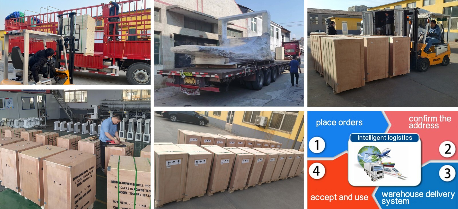
Shandong Laishi Automation Technology Co., Ltd. (hereinafter referred to as "Laishi") is composed of a technical team with more than 10 years of research and development and manufacturing experience. The registered capital of the company is 3 million yuan. We specialize in the research and development, production, and sales of various desktop hardness testers and metallographic sampling equipment, and provide professional research and sales services for physical and chemical testing instruments, laboratory equipment, analytical equipment, and automation equipment.
Packaging and Shipping
Have an inquiry or some feedbak for us? Fill out the form below to contact our team.
Related Product Recommendations
 SZM-W Computerized Continuous Zoom Stereo MicroscopeMore Details +
 JSZ8-W Trinocular Continuous Zoom Stereo MicroscopeMore Details +
 JSZ8-RS Type Weld Penetration MicroscopeMore Details +
 FZ0756U Digital Microscope All-in-One MachineMore Details +
 FBJ-700 Portable Metallographic MicroscopeMore Details +


Search Starts Here
Our Email